Most amazing microscope photos from photographers and scientists that took images of all things visible under a microscope.
The images below are from the 2017 Nikon Small World Contest. It doesn’t matter that this took place 2 years ago beacuse this kind of images they can never grow old and you can look at them all day long.

Photo by Dr. Bram van den Broek, Andriy Volkov, Dr. Kees Jalink, Dr. Reinhard Windoffer & Dr. Nicole Schwarz.
Immortalized human skin cells (HaCaT keratinocytes) expressing fluorescently tagged keratin.
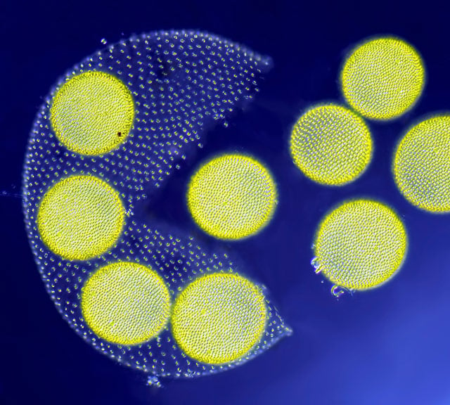
Photo by Jean-Marc Babalian.
Living Volvox algae releasing its daughter colonies.
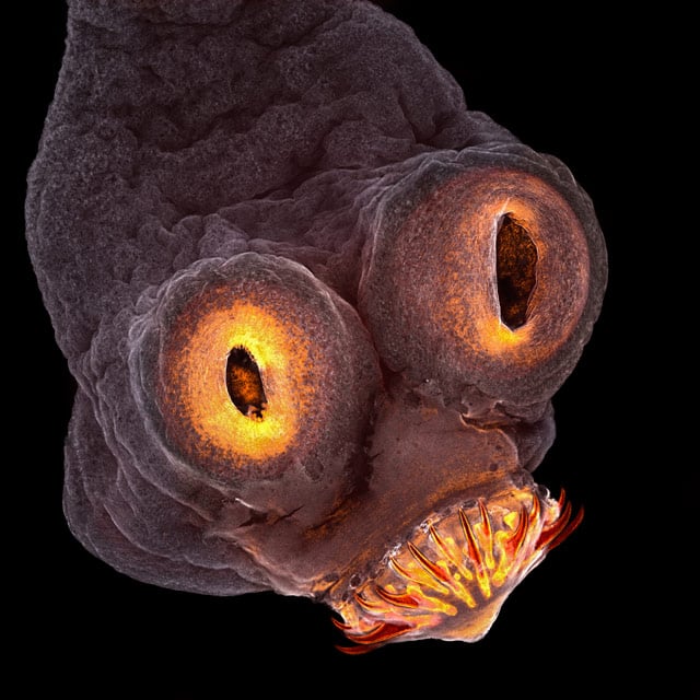
Photo by Teresa Zgoda.
Taenia solium (tapeworm) everted scolex.
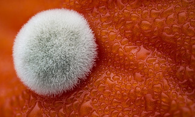
Photo by Dean Lerman.
Mold on a tomato.
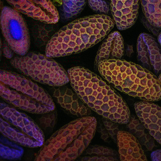
Photo by Dr. David A. Johnston.
Lily pollen.
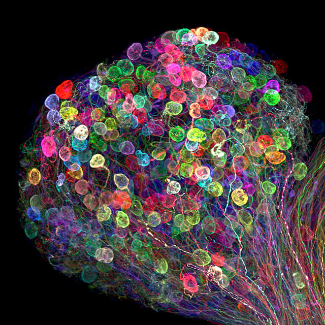
Photo by Dr. Ryo Egawa.
Individually labeled axons in an embryonic chick ciliary ganglion.
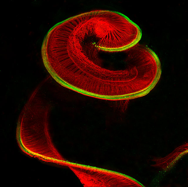
Photo by Dr. Michael Perny.
Newborn rat cochlea with sensory hair cells (green) and spiral ganglion neurons (red).
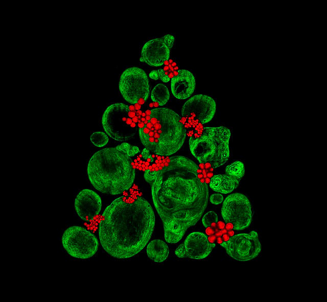
Photo by Catarina Moura, Dr. Sumeet Mahajan, Dr. Richard Oreffo & Dr. Rahul Tare.
Growing cartilage-like tissue in the lab using bone stem cells (collagen fibers in green and fat deposits in red).
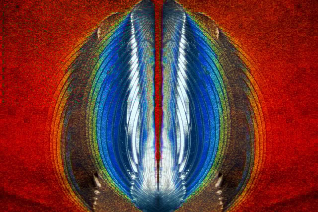
Photo by Steven Simon.
Plastic fracturing on credit card hologram.
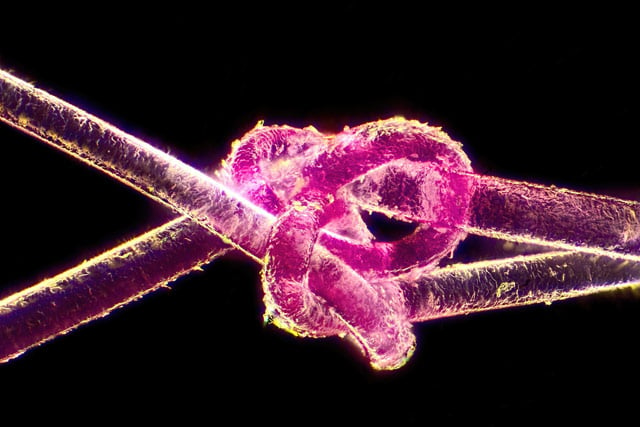
Photo by Harald K. Andersen.
Dyed human hair.
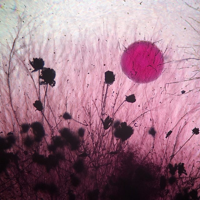
Photo by Tracy Scott.
Aspergillus flavus (fungus) and yeast colony from soil.
© 2019 Nikon Instruments Inc.